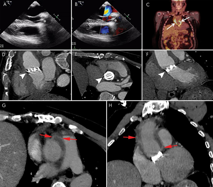
Figure 8:
Images show a 48-year-old man with a history of Bentall surgery with placement of a mechanical valve 3 years prior to presentation with fever and positive blood cultures for Staphylococcus aureus. A, B, Transesophageal echocardiography images show a normal appearance of the mechanical valve (*) with no signs of endocarditis. C, Fluorine 18 fluorodeoxyglucose (18F-FDG) PET/CT image shows intense 18F-FDG uptake around the aortic prosthesis (arrows). D–F, Contrast-enhanced CT images show the mechanical valve (arrowheads) and, G, H, fluid collection with enhancing rim surrounding the aortic graft compatible with abscess (red arrows).

