
Figure 11b:
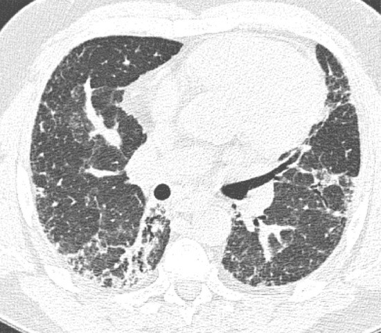
CT pattern indeterminate for usual interstitial pneumonia. (a–c) Axial inspiratory CT images demonstrate peripheral and lower lung–predominant reticular abnormality with architectural distortion, including traction bronchiectasis. However, there is slightly more ground-glass abnormality than typically seen with a confident diagnosis of a usual interstitial pneumonia pattern, and the reticular abnormality extends along the bronchovascular bundles rather than being confined to the subpleural lung.