
Figure 13b:
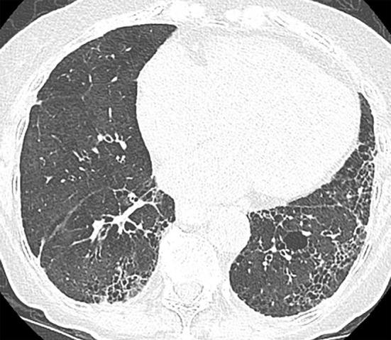
Nonspecific interstitial pneumonia. (a) Axial CT scan demonstrates fibrotic nonspecific interstitial pneumonia early in the disease course with lower lung fibrosis and ground-glass opacity with some subpleural sparing. (b) Axial CT scan 10 years later demonstrates substantial progression of the pulmonary fibrosis, which now demonstrates increased reticular abnormality, less ground-glass abnormality, and more traction bronchiectasis. Understanding the evolution of the disease by reviewing the prior imaging is critical in these cases.