
Figure 15a:
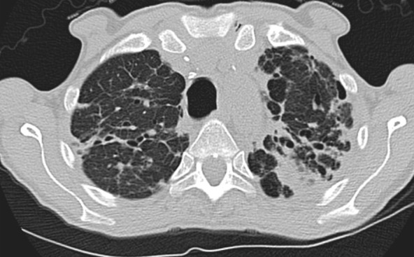
Advanced fibrotic sarcoidosis. (a–c) Axial inspiratory CT and (d) coronal inspiratory CT images demonstrate mid to upper lung–predominant pulmonary fibrosis with marked architectural distortion, associated lung nodularity, and some mosaic attenuation. Note also the calcified mediastinal lymph nodes. This case would be categorized as most consistent with a nonidiopathic pulmonary fibrosis diagnosis based on these features.