
Figure 19b:
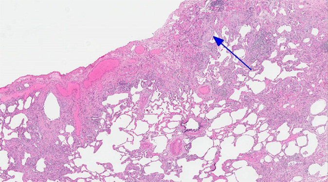
CT-histologic correlation in usual interstitial pneumonia. (a) Coronal CT image shows reticular abnormality and mild honeycombing with subpleural and lower lung predominance, typical of usual interstitial pneumonia. (b) Photomicrograph from histologic examination shows remodeling of the lung architecture by predominantly subpleural and paraseptal dense fibrosis, with scattered fibroblastic foci (blue arrow). Areas of normal lung are also seen, mainly in the centrilobular region, indicating temporal heterogeneity. (Histologic image courtesy of Rosane Duarte Achcar, MD, National Jewish Health.)