
Figure 1b:
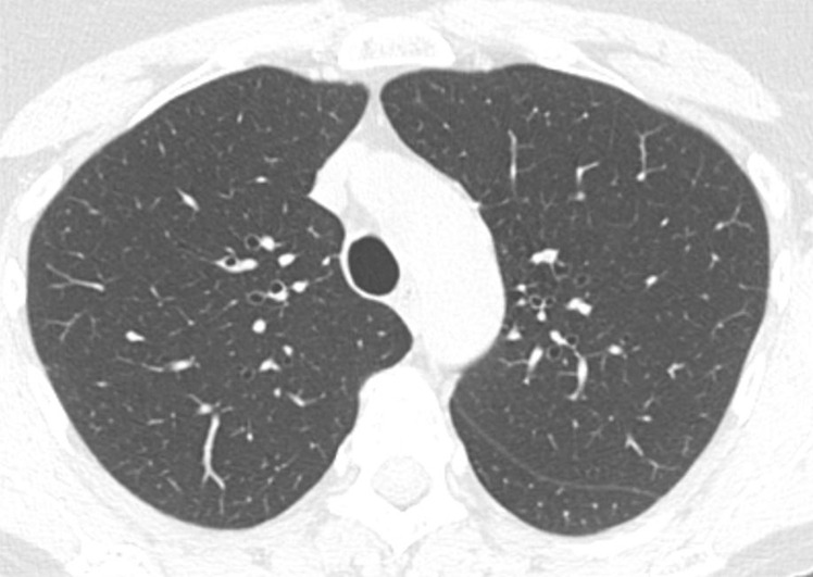
Axial thin-section CT scans in a healthy patient. (a) Inspiratory examination reconstructed with 5-mm thick sections and soft-tissue reconstruction demonstrates relative loss of definition of the interlobular septa and secondary pulmonary lobule. (b) Inspiratory sequence reconstructed with 1-mm thin sections and moderate edge-enhancing kernel reconstruction shows normal appearance. (c) Expiratory imaging shows bowing of the posterior tracheal wall and diffuse mildly heterogeneous increase in lung attenuation. Note that mild heterogeneous lung attenuation is normal.