
Figure 2:
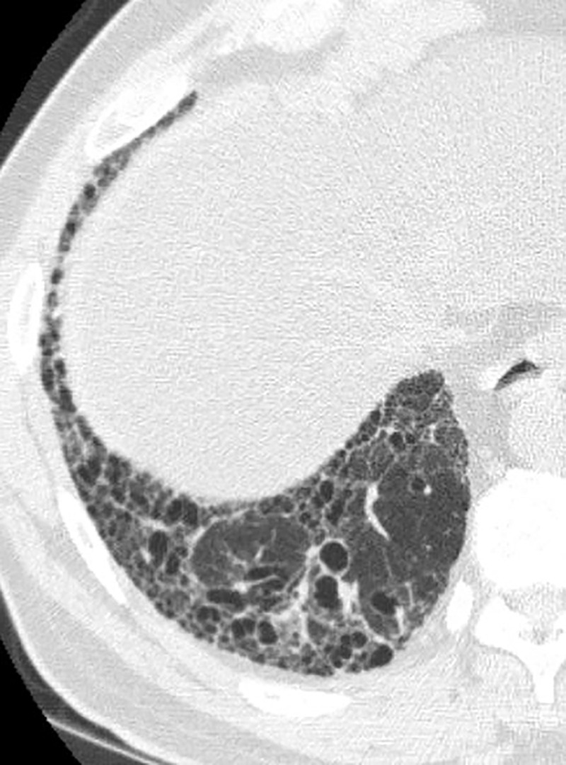
Reticular abnormality and architectural distortion. Reticular abnormality consists of a fine network or mesh of overlapping linear lines within the secondary pulmonary lobule. It is this network or mesh that separates reticular abnormality from ground-glass opacities, which are more homogeneous (see Figure 5). This case also shows associated architectural distortion with the normal secondary pulmonary lobules demonstrating tethering and warping of the normal hexagonal appearance. There is also mild honeycombing best seen in the costophrenic sulcus.