
Figure 20b:
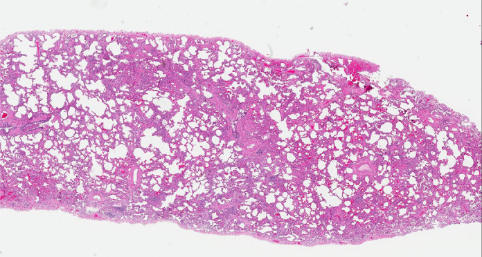
CT-histologic correlation in nonspecific interstitial pneumonia. (a) Axial CT image shows predominant ground-glass abnormality, with mild subpleural reticular abnormality. (b) Histologic image shows a diffuse homogeneous process with expansion of the alveolar septa by chronic inflammation and patchy interstitial scarring, unassociated with honeycomb change or substantial remodeling of the underlying lung architecture. In contrast to UIP, the abnormality is temporally homogeneous. (Hematoxylin-eosin stain) (Histologic image courtesy of Rosane Duarte Achcar, MD, National Jewish Health.)