
Figure 21b:
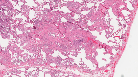
CT-histologic correlation in fibrotic hypersensitivity pneumonitis (HP). (a) Coronal CT image shows lower lung–predominant reticular and ground-glass abnormality. The findings of mosaic attenuation and peribronchovascular predominance are highly suggestive of fibrotic HP. (b) Photomicrograph from histologic examination shows centrilobular scarring compatible with fibrotic HP. (Hematoxylin-eosin stain) (c) Peribronchiolar poorly formed nonnecrotizing granulomas are also present, highly suggestive of HP. (Hematoxylin-eosin stain) (Histologic images courtesy of Rosane Duarte Achcar, MD, National Jewish Health.)