
Figure 4a:
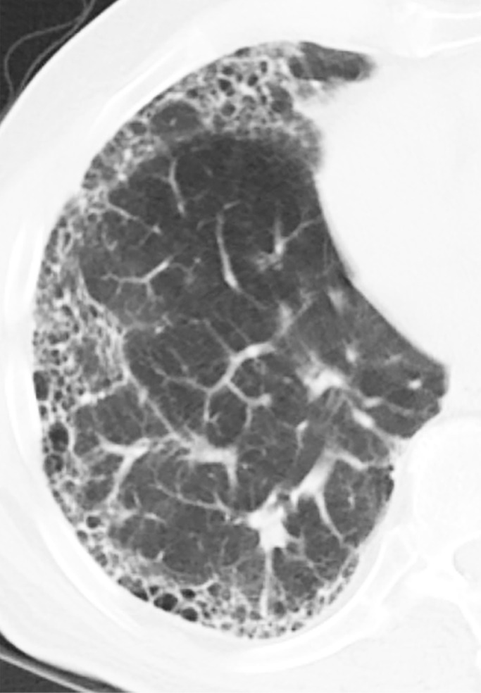
Honeycomb cysts and paraseptal emphysema. (a) Zoomed view of axial CT demonstrates classic honeycomb cyst formation with stacking subpleural cysts and associated adjacent fibrosis in a patient with idiopathic pulmonary fibrosis. (b) Zoomed view of axial CT in a different patient demonstrates paraseptal emphysema, characterized by a single layer of subpleural cysts, without any evidence of associated fibrosis in a patient with a history of marijuana cigarette smoking. The cysts of paraseptal emphysema are typically more than 1.0 cm in diameter, larger than honeycomb cysts, and have thinner walls.