
Figure 5:
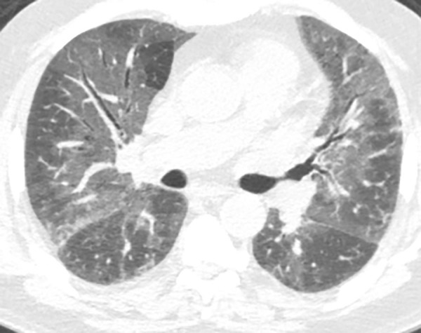
Ground-glass opacities. Axial CT image demonstrates homogeneous areas of increased lung attenuation in which the increased opacity does not obscure the underlying bronchial and vascular structures. This finding is not a feature typical of usual interstitial pneumonia or idiopathic pulmonary fibrosis but can be seen in the setting of overlapping or superimposed disease.