
Figure 6:
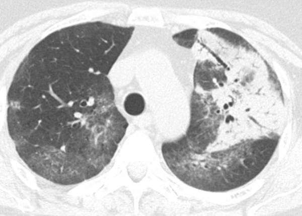
Consolidation. Left upper lobe consolidation in a patient with infectious pneumonia. The lung attenuation is diffusely increased, with air bronchograms, and the underlying vessels are completely obscured by the airspace opacity. Ground-glass opacities are seen in the nonconsolidated lung bilaterally. This finding is not a feature typical of usual interstitial pneumonia or idiopathic pulmonary fibrosis but can be seen in the setting of overlapping or superimposed disease.