
Figure 8b:
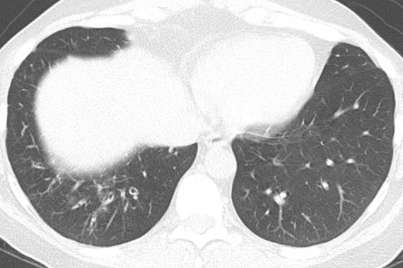
Comparison of traction bronchiectasis with bronchiectasis from chronic airway inflammation. (a) Axial CT scan demonstrates traction bronchiectasis in a patient with fibrotic nonspecific interstitial pneumonia. Note the substantial reticular abnormality and architectural distortion adjacent to the irregular dilated airways. (b) Axial CT scan demonstrates cylindrical bronchiectasis in the right lower lobe of a patient with underlying immunodeficiency. In contrast to traction bronchiectasis, this postinflammatory bronchiectasis lacks adjacent fibrosis and “tethering” of the airways. There are also associated findings of bronchial wall thickening and mucoid impaction.