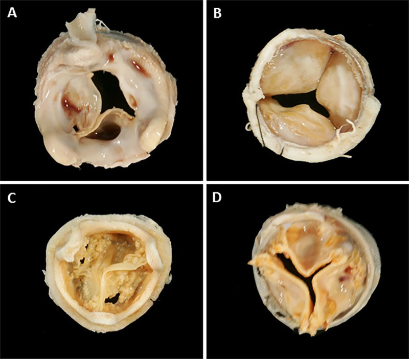
Figure 2:

Images show examples of features of bioprosthetic heart valves (BPHVs) pathology seen at gross examination at explant. BPHV explants demonstrate, A, large amounts of fibrosis with areas of thrombus on aortic aspect of surgical heart valve (SHV) with pericardial leaflets; B, fibrosis on ventricular aspect on SHV with pericardial leaflets; and calcified SHVs with, C, leaflet tears in valves with porcine and, D, pericardial leaflets.
