editorial
. 2020 Jan 16;2(1):e190153. doi: 10.1148/ryct.2019190153

Figure 1:

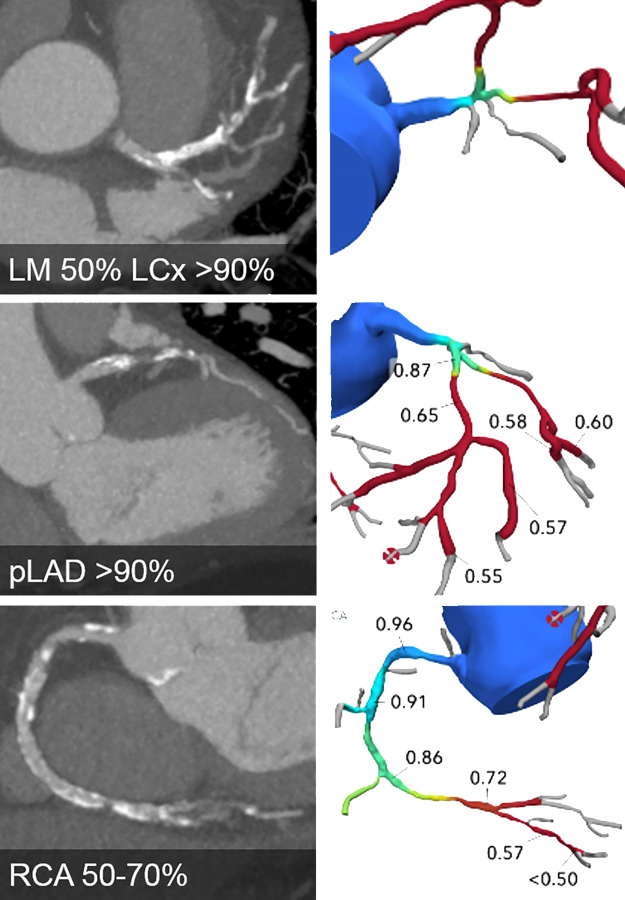
CT angiographic images (left) and FFR CT models (right) show severe three-vessel disease with anatomically and functionally significant obstructive disease of the left main (LM), left anterior descending (LAD), left circumflex (LCx), and right coronary artery (RCA) (white arrowheads). Future developments may result in such patients being triaged straight to coronary artery bypass graft surgery without the need for further testing.

CT angiographic images (left) and FFR CT models (right) show severe three-vessel disease with anatomically and functionally significant obstructive disease of the left main (LM), left anterior descending (LAD), left circumflex (LCx), and right coronary artery (RCA) (white arrowheads). Future developments may result in such patients being triaged straight to coronary artery bypass graft surgery without the need for further testing.