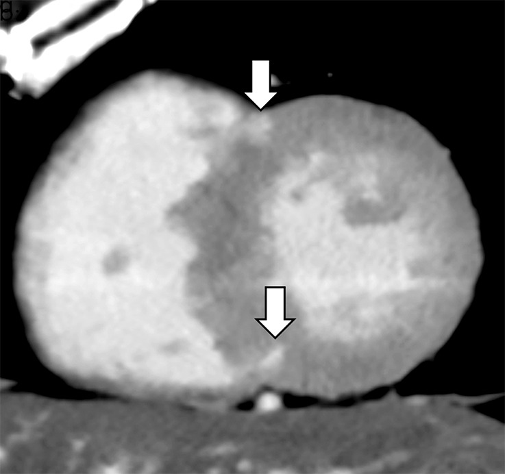
Figure 2b:

Images show comparison of (a–c) late iodine enhancement (LIE) CT images and (d) late gadolinium enhancement (LGE) cardiac MR image in a 63-year-old woman with hypertrophic cardiomyopathy. (a) Standard 120-kVp image, (b) virtual monochromatic image at 50 keV, and (c) iodine density image acquired by using dual-energy LIE CT. (d) LGE cardiac MR image shows LGE confined to anterior and posterior right ventricular insertion points. Late-enhancing lesions were more clearly visualized on virtual monochromatic image at 50 keV and iodine density image than on standard 120-kVp image.
