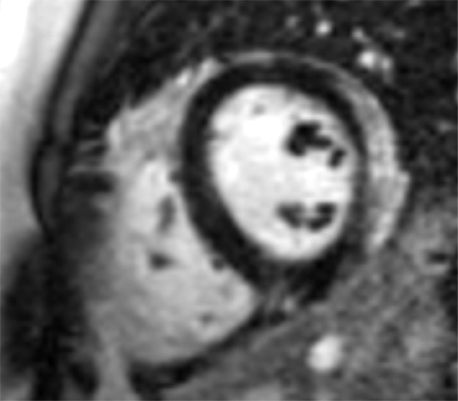
Figure 3a:

Images in a 24-year-old male patient with acute myocarditis. (a) Late gadolinium enhancement (LGE) cardiac MR image in the short-axis view. (b) Two-dimensional region-of-interest segmentation of the LGE area. (c) Three-dimensional volume-of-interest segmentation of the LGE area.
