
Fig 2.
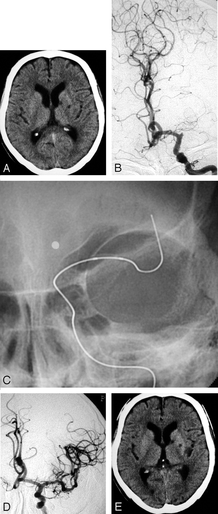
A 76-year-old man (case 2).
A, A CT on admission shows no LDA, without small lacunar infarction in the left basal ganglia. B, A preprocedural angiogram shows an occlusion of the left MCA trunk. C, A deflated microballoon is gently advanced to the embolus. D, An angiogram obtained immediately after the treatment shows complete recanalization. E, On the CT scan obtained 24 hours after treatment, there is no change compared with the initial CT on admission.