
Fig 4.
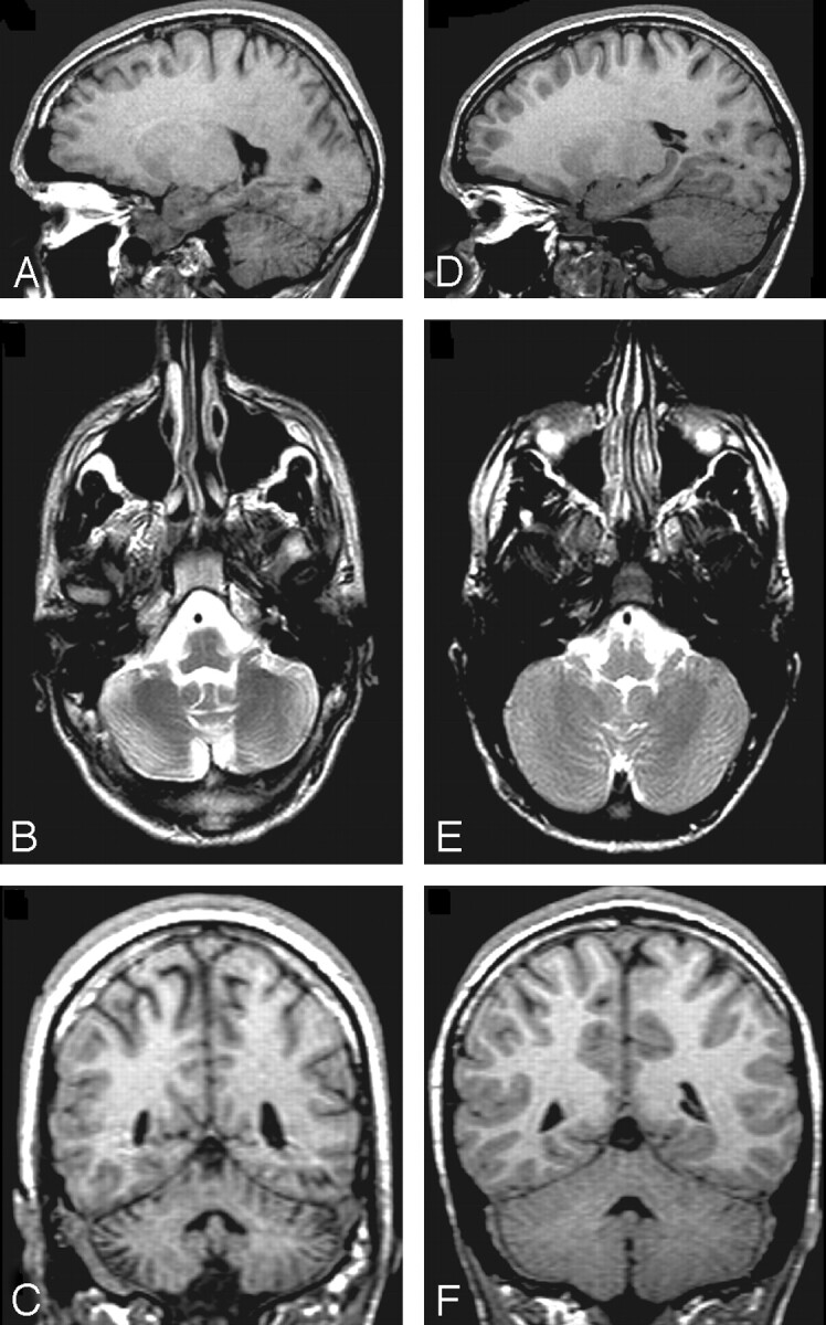
T1WI sagittal (A), T2WI axial (B), and T1WI coronal (C) images of the cerebellum from a 16-year-old boy with TBI (initial GCS = 3) and a TD child of comparable age and sex (D–F). The child with TBI has diffuse cerebellar atrophy and increased CSF within the cerebellar folia that can be noted in all planes.