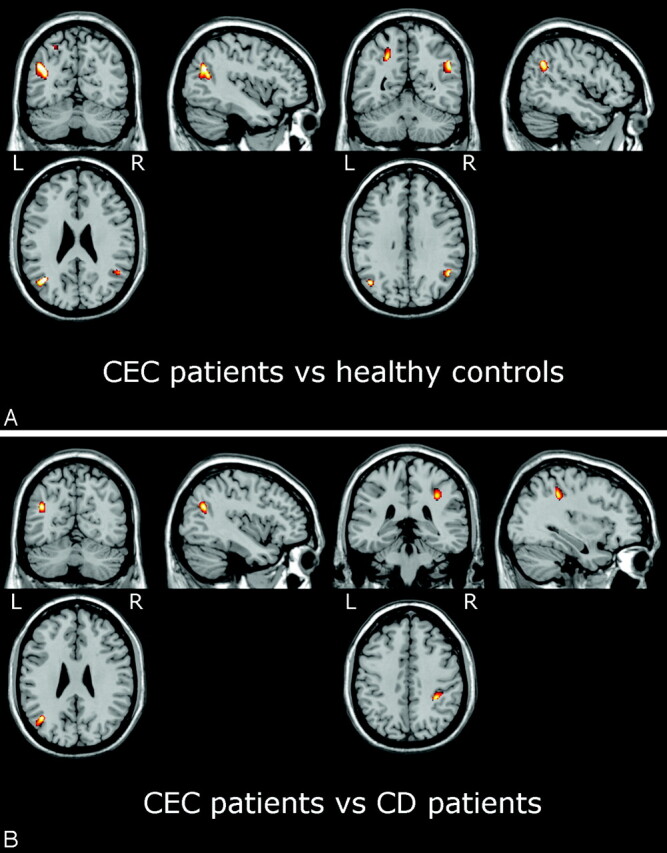
Fig 3.

SPM2 results showing areas of decreased MTR in patients with CEC versus healthy control subjects (A) and versus patients with CD (B) superimposed on anatomic references. In patients with CEC, areas of significantly (PFDRcorr<0.05) decreased MTR compared with control subjects (A) include the white matter of the parietal lobe bilaterally (see Table 3; unthresholded statistical map is provided as supplemental Fig 2). Compared with patients with CD (B), the patients with CEC show areas of decreased MTR (Puncorr < 0.001) in the white matter of the left temporal lobe and right parietal lobe (see Table 3).
