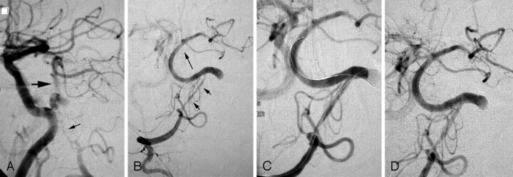
Fig 3.
A, 66-year-old man with recurrent episodes of dizziness, nausea, and diplopia. Lateral view of arteriogram performed with the catheter in the innominate artery. The right vertebral artery has a hairlike residual lumen beyond the posterior inferior cerebellar artery (small arrow); the basilar artery is supplied in large part from the anterior circulation via the posterior communicating artery (larger arrow). The dominant left vertebral is occluded just above the posterior inferior cerebellar artery. B, After initial angioplasty with a 1.5-mm balloon, distal perfusion is improved (long arrow indicating antegrade flow in the basilar artery), but a severely stenotic lumen persists (short arrows). C, After placement of overlapping stents in the distal vertebral artery, there is antegrade filling of both posterior cerebral arteries indicating increased perfusion. D, Initial follow-up examination at 6 weeks reveals continued patency. The patient was followed up with CT angiography, and the stents remain patent 2 and a half years later.

