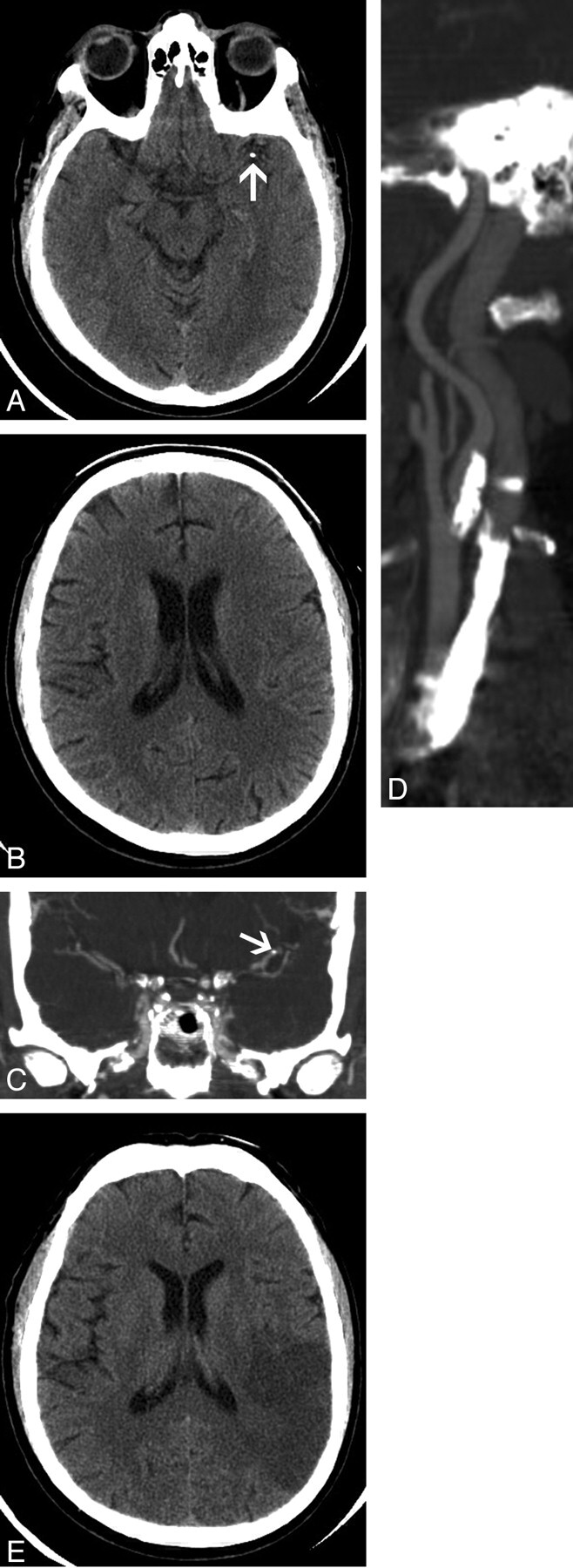
Fig 1.

A 66-year-old man with a calcified cerebral embolus to the left middle cerebral artery.
A, Axial 2.5-mm image from a noncontrast brain CT scan shows a calcified embolus in the distal M1 segment of the left middle cerebral artery (arrow).
B, Axial 2.5-mm image obtained at a higher level shows a normal left parietal lobe, with normal gray-white matter differentiation.
C, Coronal image from a CT angiogram from the level of the aortic arch to the circle of Willis shows calcified embolus in the distal M1 segment of the left middle cerebral artery (arrow).
D, Coronal oblique image from the same examination as C shows high-grade stenosis of the left internal carotid artery with extensive calcified plaque.
E, Follow-up noncontrast brain CT scan, obtained 1 day after A and B, shows an acute infarct in the left middle cerebral artery territory with loss of normal gray-white matter differentiation and local mass effect.
