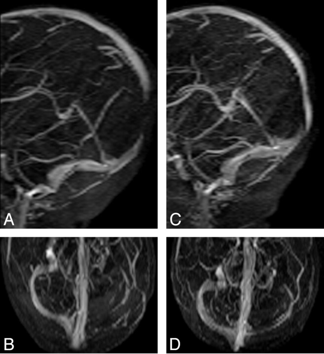
Fig 4.
A and B, MIP images of coronal 2D TOF MRV acquired with TE of 5.5 ms demonstrate flow gap in the superior sagittal sinus and left transverse sinus. The flow gap is artifactual as a result of in-plane saturation. C and D, MIP images of coronal 2D TOF MRV acquired with TE of 4.9 ms. Flow gap due to in-plane saturation is minimized by using shorter TE (4.9 ms) and thereby improves visualization of superior sagittal and transverse sinus. The left transverse sinus is smaller compared with the right transverse sinus but patent, possibly due to congenitally smaller caliber left transverse sinus.

