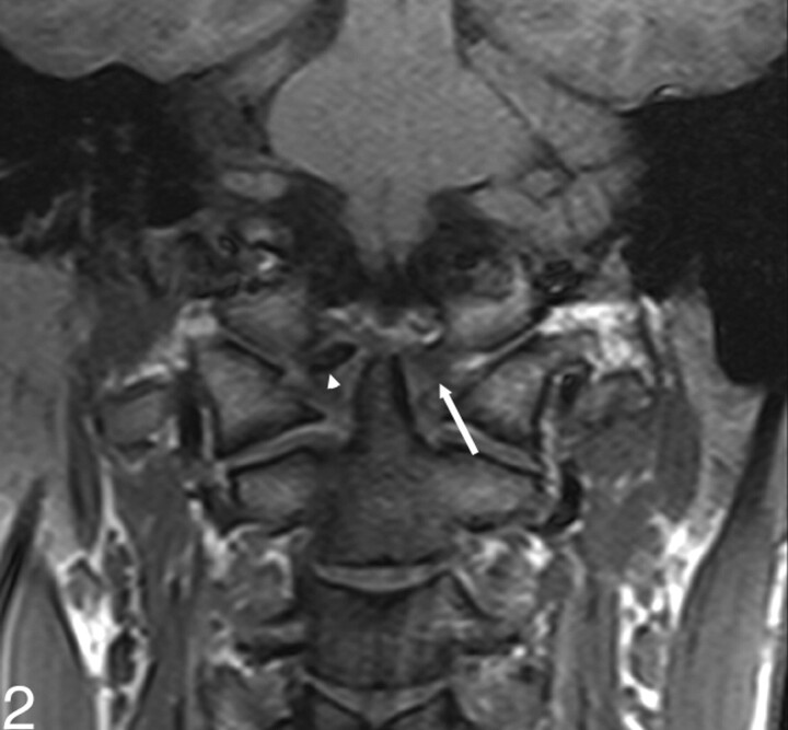
Fig 2.
MR image with coronal T1-weighted sequences shows a nodular calcific attenuation (arrowhead) in the lateral part of the right side alar ligament. It expands in thickness and width beyond the anatomic boundaries of alar ligament. The left alar ligament (white arrow) is a well-defined structure running caudocranially from the apex of odontoid to the occipital condyle, with intermediate signal intensity without any pathologic change.

