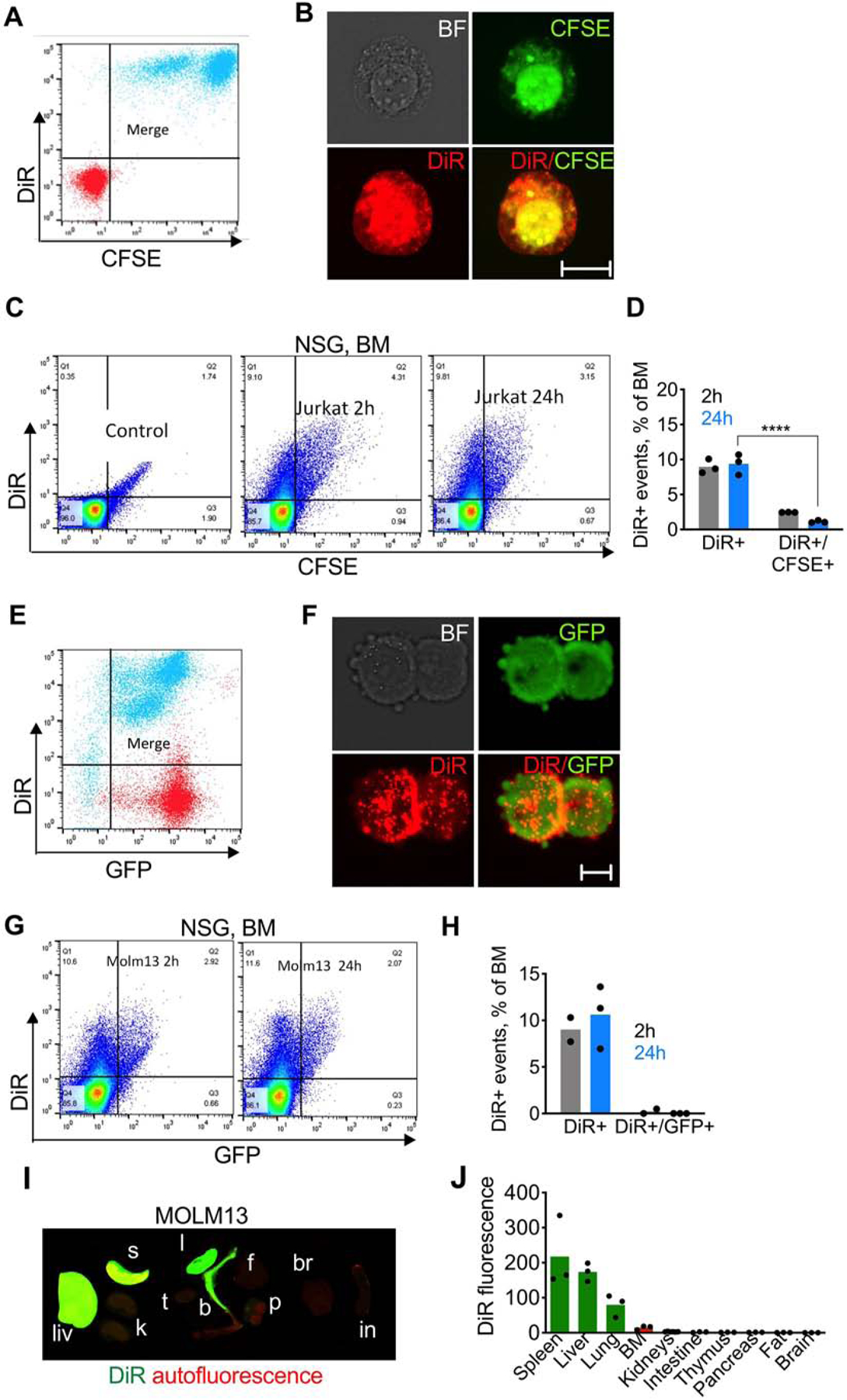
Fig. 2. ICLs accumulate in the BM independently of the injected cells:

A-B) DiR and CFSE dual labeling of Jurkat cells. Red, before labeling; cyan, after labeling; C-D) flow cytometry of BM shows high percentage of DiR+ events but low percentage of DiR+/GFP+ events (n=3; 2-way ANOVA with multiple comparisons). Control is the BM of non-injected mice; E-F) DiR labeling of GFP-MOLM13 cells. Red, before labeling; cyan, after labeling; G-H) flow cytometry of BM after injection of DiR/GFP-MOLM13 shows high percentage of DiR+ events but almost no DiR+/GFP+ events; I) ex vivo NIR imaging of organs of NSG mice injected with DiR/GFP-MOLM13; J) image quantification.
