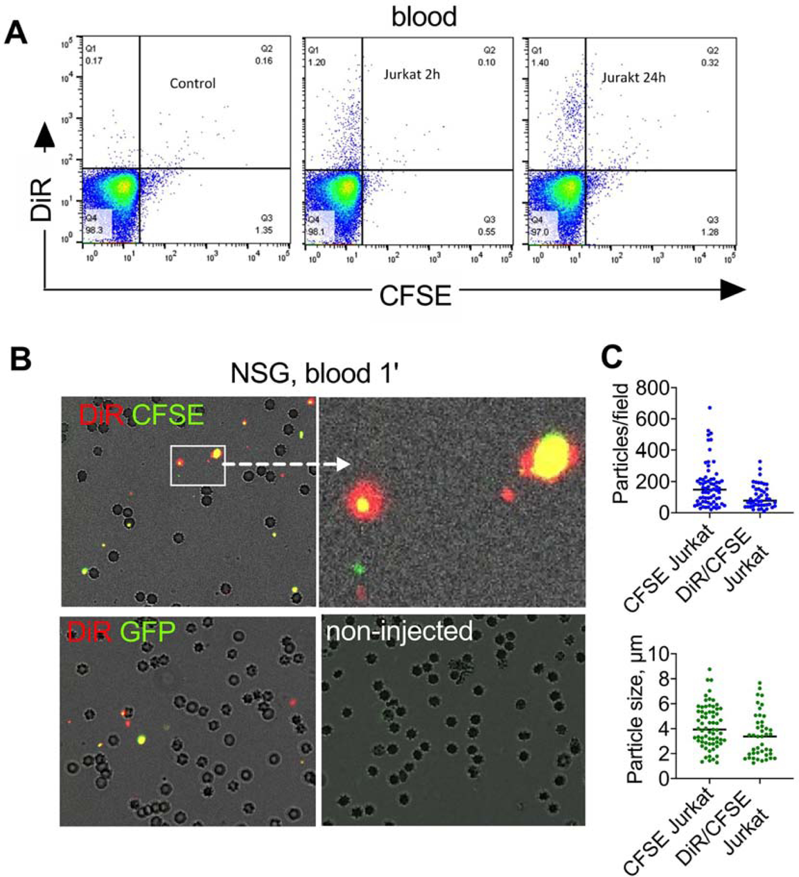
Fig. 5. Cells release microparticles in vivo:

A) Flow cytometry of peripheral blood (RBCs lysed) after injection DiR/CFSE Jurkat cells in NSG mice shows no cells but some DiR+ events; B) microparticles in blood 1 min post-injection of DiR/CFSE Jurkat (top left and center) and DiR/GFP-MOLM13 (top right). Many RBCs are visible. Some of the particles are as large as RBCs, but smaller than intact cells. Fluorescence was enhanced to demonstrate colocalization; graphs show quantification of particles in blood 1 min after injection of DiR/CFSE Jurkat or CFSE Jurkat (n=2 mice, at least 40 microscopic fields counted). Particles were also observed after injection of non-labeled GFP-MOLM13 and DiR/CFSE splenocytes (supplemental data); C) low magnification images of freshly excised organs of NSG mice injected with DiR/CFSE Jurkat (2h post injection) show accumulation of CFSE and DiR in the lungs, but preferential accumulation of DiR in the liver and spleen. Size bar, 100 µm.
