. 2021 Feb 25;3(1):e200512. doi: 10.1148/ryct.2021200512

Figure 2:

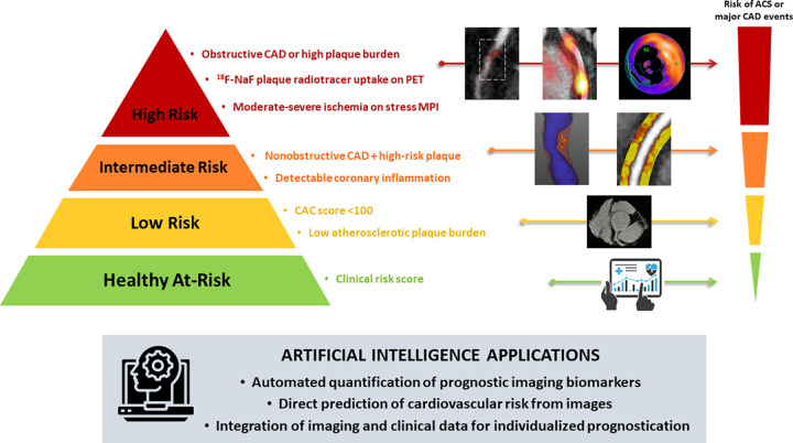
Artificial intelligence (AI) in cardiovascular imaging for risk stratification. Clinical risk stratification based on anatomic and functional imaging assessment of coronary artery disease (CAD). The image case examples (from left to right) correspond to the descriptions (from top to bottom) adjacent to the risk pyramid. AI algorithms can perform automated measurements of prognostic biomarkers from image data. Additionally, conventional or AI-based imaging parameters can be combined with clinical data using machine learning models for individualized risk prediction. ACS = acute coronary syndrome, CAC = coronary artery calcium, 18F-NaF = fluorine 18 sodium fluoride, MPI = myocardial perfusion imaging.

Artificial intelligence (AI) in cardiovascular imaging for risk stratification. Clinical risk stratification based on anatomic and functional imaging assessment of coronary artery disease (CAD). The image case examples (from left to right) correspond to the descriptions (from top to bottom) adjacent to the risk pyramid. AI algorithms can perform automated measurements of prognostic biomarkers from image data. Additionally, conventional or AI-based imaging parameters can be combined with clinical data using machine learning models for individualized risk prediction. ACS = acute coronary syndrome, CAC = coronary artery calcium, 18F-NaF = fluorine 18 sodium fluoride, MPI = myocardial perfusion imaging.