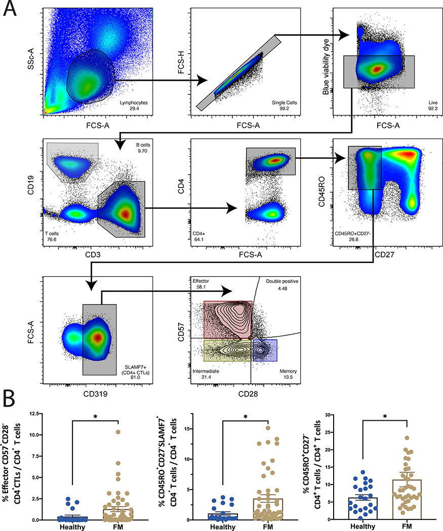
Figure 1. CD4+CTLs are expanded in fibrosing mediastinitis.
(A) Flow cytometry gating strategy to identify effector CD4+ T cells (CD4+CD45Ro+CD27-), CD4+CTLs (CD4+CD45Ro+CD27-SLAMF7+), and effector CD4+CTLs (CD4+CD45Ro+CD27-SLAMF7+CD28loCD57hi) (top). (B) Comparison of the frequency of effector CD4+ T cells (right), CD4+CTLs (center), and effector CD4+CTLs (left). Quantified as % of total CD4+ T cells and expressed as means +/− SE (Control n=21, FM n= 47). *P<0.05 by two-tailed Mann-Whitney t-test.

