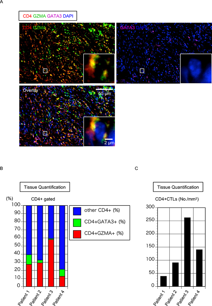
Figure 3. CD4+CTLs are an important part of the CD4+ T cells infiltrating tissue from patient with FM.
(A) Representative multicolor immunofluorescence image of cells co-expressing CD4 (red) and Granzyme A (GZMA)(green) that infiltrate the mediastinum in FM. (B) Relative proportions of GZMA+CD4+CTLs, GATA3+TH2 cells and other cells (Each column represents a patient; n = 4). (C) Absolute number of GZMA+CD4+CTLs in mediastinal biopsy of 4 patients with FM.
Nuclei were stained with DAPI (Blue) CD4 was in red, GZMA was green and GATA3 was purple. The inset shows a single CD4+CTL with extranuclear CD4 and GZMA.

