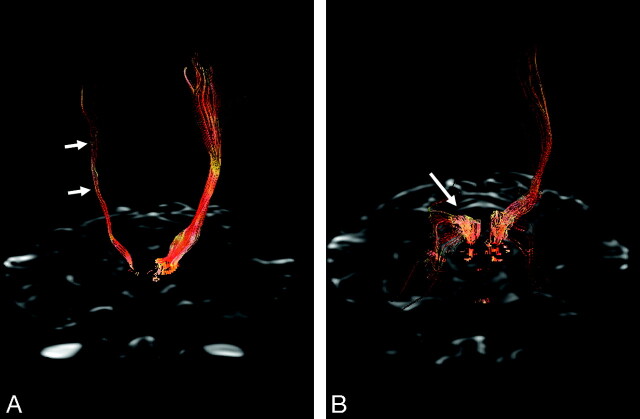
Fig 2.
3D fiber tractography of the pyramidal tracts (A) and sensory tract through the dorsal pons and thalamus (B). A, The pyramidal tracts are clearly depicted on the left hemisphere but are sparse on the right hemisphere (arrows). B, The sensory tract on the right side is prominently involved (arrow).

