Fig 2.
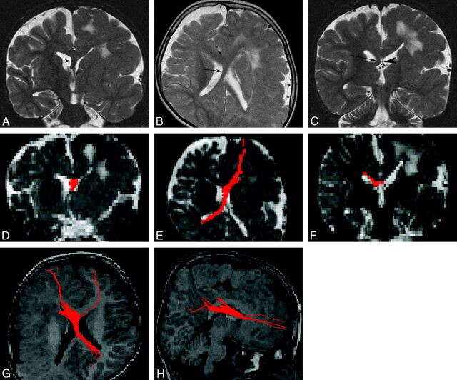
An 8-year-old boy with left hemimegalencephaly (case 1). A, Coronal T2-weighted inversion recovery image shows a white matter–intensity structure between the 2 anterior horns (arrow). B, Axial T2-weighted image shows a midsagittal bandlike structure between the bodies of the 2 lateral ventricles (arrow). It appears to cross from anterior-left to posterior-right. C, Coronal T2-weighted inversion recovery image. The left fornix (arrowhead) is normal, while the right one (arrow) is thick. D–F, 2D coronal (D), 2D axial (E), and 2D coronal (F) views on fiber tract (FT) reconstruction. The images correspond to A, B, and C, respectively. Aberrant midsagittal fibers penetrate the midsagittal bandlike structures shown in A–C. G and H, 3D superior (G) and 3D right lateral (H) views on FT reconstruction. The fibers run between the 2 frontal lobes and the contralateral occipital and parietal lobes. The main fibers pass through the right crus of the fornix.

