Fig 3.
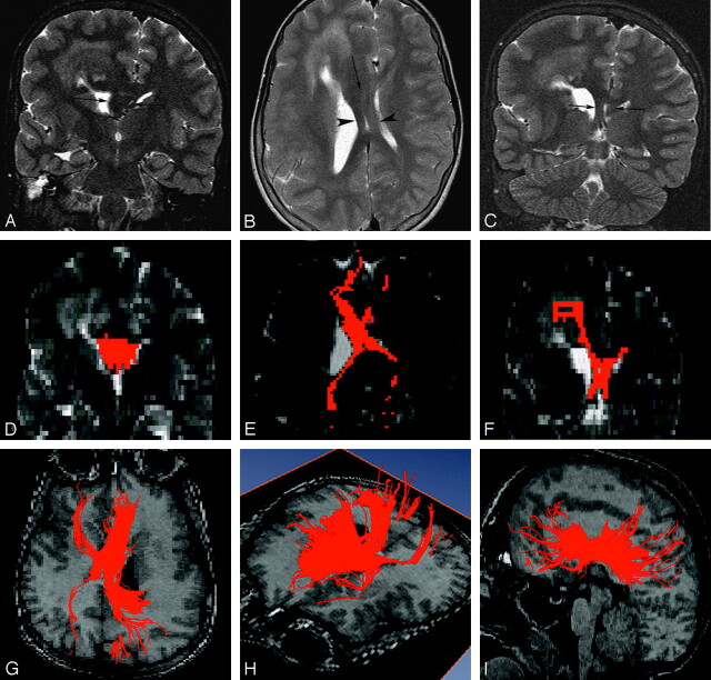
An 11-year-old boy with right hemimegalencephaly (case 2). A, Coronal T2-weighted inversion recovery image shows markedly increased width between the 2 anterior horns occupied by a white matter–intensity structure (arrows). B, Axial T2-weighted image demonstrates an abnormal white matter–intensity bandlike structure between the 2 lateral ventricles (arrow), which separates in the posterior portion and runs a course similar to that of the cruces of the fornices (arrowheads). C, Coronal T2-weighted inversion recovery image posterior to that in A also detects bilateral marked thickening of the cruces of the fornices (arrows). D–F, 2D coronal (D), 2D axial (E), and 2D coronal (F) views on FT reconstruction. These images correspond to A, B, and C, respectively. Aberrant midsagittal fibers penetrate the midsagittal abnormal white matter–intensity structures shown in A–C. G–I, 3D superior (G), 3D left lateral superior (H), and 3D left lateral (I) views on FT reconstruction. Aberrant massive fibers connect the bilateral frontal and occipital and parietal lobes passing through abnormal midsagittal structures. They are dominant ipsilaterally.

