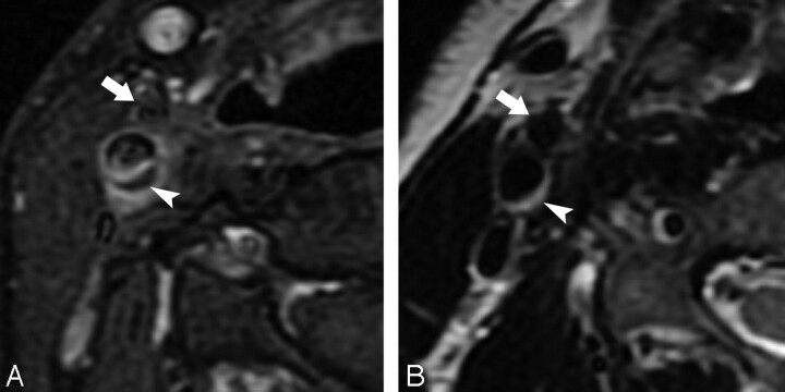
Fig 3.
Right ICA at presentation (A) and at 5 months (B). A, Axial fat-suppressed postcontrast T1-weighted image demonstrates enhancement of the ICA wall and a fatty plaque (arrowhead) deep to the intima. Arrow indicates right external carotid artery (ECA). B, Axial fat-suppressed T1-weighted image demonstrates resolution of mural thickening and high signal intensity; disappearance of the fatty plaque with irregular mural thickening is now seen (arrowhead), consistent with scarring or residual inflammatory tissue. Arrow indicates right ECA.

