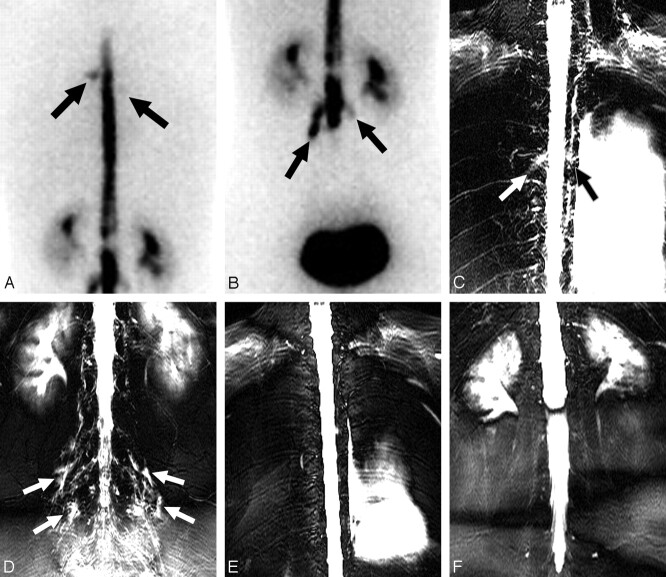
Fig 3.
A 33-year-old woman with spinal CSF leak syndrome and multiple CSF leaks in the bilateral thoracic and lumbar spine. A and B, Posterior projection of RIC shows diffusion of the radioisotope into the extra-arachnoidal space in the region of the upper thoracic spine (arrows), predominantly on the left side (A) and in the lumbar spine (B). C and D, MRM shows hyperintensities along multiple nerve root sleeves in the upper thoracic spine (arrows, C) and in the lumbar spine (arrows, D). E and F, On 1-month follow-up MRM, multiple hyperintensities around the nerve root sleeves disappeared.

