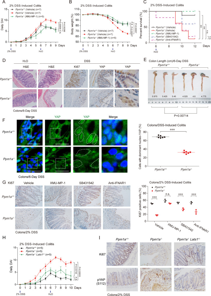
Fig 6. PPM1A is indispensable for murine intestinal regeneration upon colitis.
(A, B) DSS (2%)-induced colitis was performed in wild-type and PPM1A KO mice at 10–12 weeks age. More severe phenotypes of daily DAI (A) and body weight loss (B) were detected in PPM1A KO mice, starting at Day 6. Administration of XMU-MP-1, a small molecular inhibitor of MST kinase, largely restored the colitis phenotypes in Ppm1a−/− mice (A and B). (C) DSS-induced colitis resulted in the substantial death of Ppm1a−/− mice, during the extended observation and by log-rank test in statistics. Administration of XMU-MP-1, but not the inhibitors/neutralizing antibody for TGF-β and IFN-I signaling, survived Ppm1a−/− mice. (D) DSS treatment led to a severe structural loss of intestine villus, which was largely recovered through intestine regeneration in wild-type mice, as examined at Day 8 (DSS induction for 6 days). The recovery of intestine in Ppm1a−/− mice was markedly compromised, as evidenced by the disorganized intestine villi (second panel, H&E staining) and the obviously decreased number of proliferating cells (Ki67 positive) (third panel, Ki67 IHC). A compromised level of the neucleo-YAP was also seen in Ppm1a−/− intestine (fourth and fifth panels, YAP IHC). (E) The length of mice colons post DSS treatment indicated a more severe impairment of intestinal regeneration and/or inflammatory responses in Ppm1a−/− mice at Day 8 (DSS induction for 6 days). (F) Immunofluorescence imaging showed a dramatic difference of YAP cellular distribution in intestine villi from wild-type and PPM1A KO mice. A substantial decrease of the nucleo-YAP was seen in intestines from Ppm1a−/− mice. (G) Administration with XMU-MP-1 (1 mg/kg, once a day) via IP injection restored both the villi structure (left panel) and percentage of the proliferating cells (right panel) in Ppm1a−/− intestines. Administration of SB431542 (4.2 mg/kg, once a day) alleviated these intestine phenotypes but anti-IFNAR1 (0.2 mg/mouse, once every 2 days) not. (H, I) Genetic deficiency of LATS1 (Lats1+/−) largely restored the severe colitis phenotypes upon PPM1A deficiency, as indicated by the alleviated level of disease DAI (H) and villi structure that was mostly intact (I). An increase of phospho-YAP (S112) (equivalent human S127) was seen in the absence of PPM1A, which was down-regulated by LATS1 deficiency (I). Unprocessed images of blots are shown in S1 Raw Images. Statistics source data are provided in S1 Data. DAI, disease activity index; DSS, dextran sulphate sodium; H&E, hematoxylin and eosin; IFN-I, type I interferon; IHC, immunohistochemistry; IP, intraperitoneal; KO, knockout; LATS1, large tumor suppressor kinase 1; MST, mammalian sterile 20-like kinase; phospho-YAP, phosphorylating forms of YAP; PPM1A, protein phosphatase magnesium-dependent 1A; TGF-β, transforming growth factor beta; YAP, Yes-associated protein.

