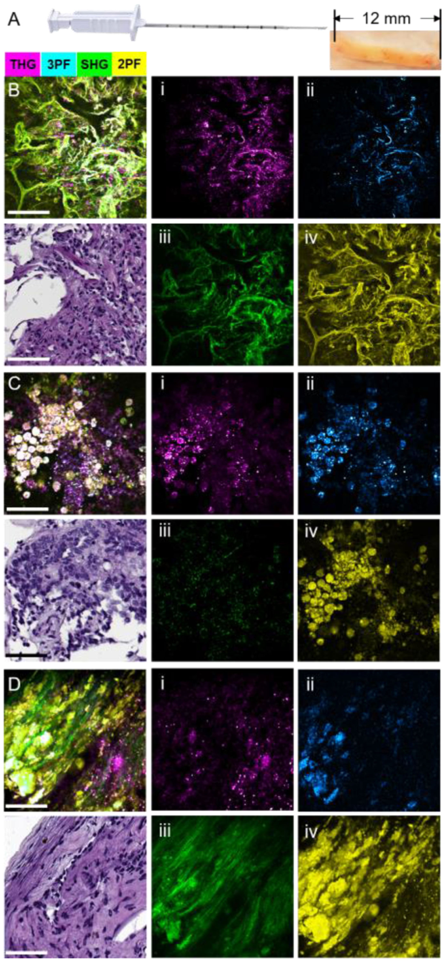
Fig. 10.

Label-free multimodal NLOI images of core NB specimens. (A) Photograph of the core NB device and representative NB specimen. NB core specimens were around 1.6-mm in diameter and 12-mm in length. (B)–(D) Intraoperative multimodal NLOI results of the tumor microenvironment from NB core specimens acquired from a canine pulmonary adenocarcinoma, showing the composite (merged) image and corresponding H&E histology, as well as each of the 4 modality channels shown separately at the right side (i)–(iv). (B) Image set from a core NB specimen acquired in vivo from a visually normal region adjacent to the tumor. (C) Image set from a core NB specimen acquired in vivo from the tumor. (D) Image set from a core NB specimen acquired ex vivo from the freshly excised lung tumor. (i) THG, magenta; (ii) 3PF, cyan; (iii) SHG, green; (iv) 2PF, yellow. Color scale is given on the top of the Figure. Scale bar represents 100 μm.
