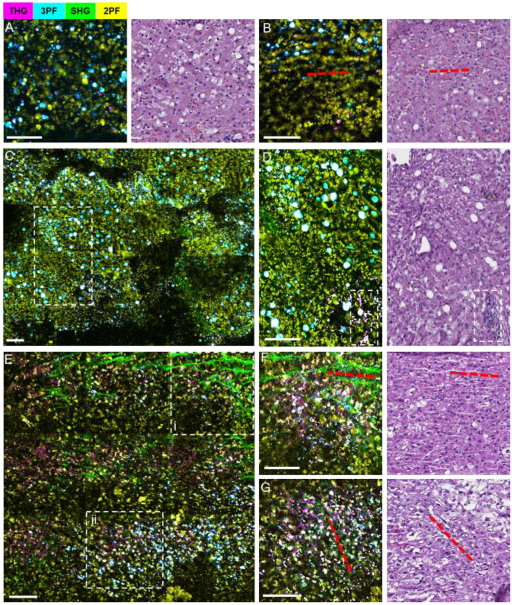
Fig. 11.

Label-free intraoperative multimodal NLOI imaging of canine liver NB cores with corresponding histology. (A), (B) Normal liver tissue biopsied adjacent to the tumor, showing sparsely distributed hepatocytes, adipocytes, and sinusoids (aligned with red dashed lines). (C) A mosaicked NLOI image from the tumor margin, with an enlarged region (dashed white box) shown in (D) where the incomplete destruction of sinusoids with fibrotic stroma content, adipocytes, and high-density foci of tumor cells (dashed box) are observed. (E) A mosaicked NLOI image of the liver tumor with regions (i, ii) enlarged in (F), (G), respectively. Red dashed lines in (F), (G) and the corresponding histology indicate general collagen fiber orientation, with dense tumor cell infiltration. Color scale is given on the top of the Figure. Scale bar represents 100 μm.
