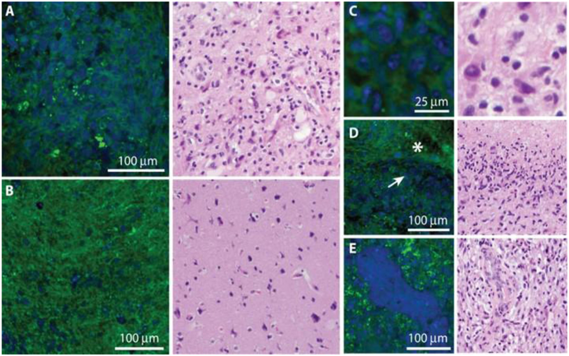
Fig. 2.

SRS microscopy of freshly excised human brain tumor specimens and corresponding H&E histology. (A) Images of viable tumor regions of show hypercellularity, in contrast with (B) normocellular regions of adjacent brain with minimal tumor infiltration. (C)–(E) Higher-magnification images of key diagnostic features of glioblastoma including cellular pleomorphism (C), pseudo-palisading necrosis, where densely cellular regions (arrow) border bland, acellular regions of necrosis (asterisk) (D), and microvascular proliferation (E). Proteins are false-colored in blue and lipids in green. Figure reprinted with permission from [31].
