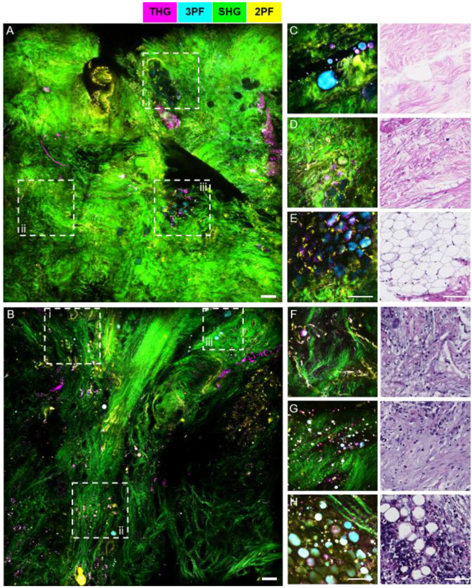
Fig. 9.

Comparison between SLAM microscopy and intraoperative NLOI systems for imaging excised human breast tissue from the same patient. (A)–(B) Large FOV SLAM images of (A) adjacent normal tissue and (B) tumor tissue excised from a breast cancer patient diagnosed with IDC. (C)–(E) images of adjacent normal breast tissue from the same patient acquired by the intraoperative NLOI system (left) and correlated with histology results (right). (F)–(H) Images of tumor tissue from the same human breast cancer patient acquired by the intraoperative NLOI system (left) and correlated with histology results (right). The dashed boxes (i–iii) in the SLAM images (A), (B) highlight features which can also be identified in the intraoperative NLOI results (C)–(E), (F)–(H). Color scale is shown on the top of the Figure. Scale bar represents 100 μm.
