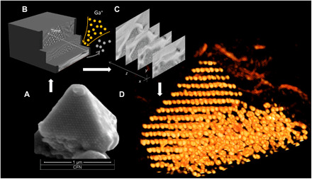
Fig. 6. 3D imaging of the silicated nanoparticle lattice using a focused ion beam processing and serial reconstruction.

(A) SEM image of a DNA-NP superlattice structure. (B) Representation of the FIB/SEM collection process wherein a 10-nm layer of sample was removed sequentially by ion milling. (C) Slices of SEM images showing the evolution of pyramid domain. (D) 3D representation of AuNP lattice showing ordered and disordered domains within the assembled volume (see also supplementary movies and the Supplementary Materials).
