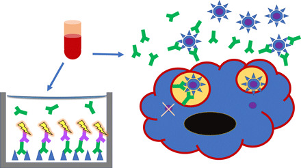
Fig. 1. The measurement of antibody binding and virus neutralization in vitro.

Blood samples are obtained from patients or experimental animals and serum is separated. (Left) Serum antibody binding is usually measured by ELISA: S proteins (blue triangles) or RBDs are immobilized in wells, S-specific antibodies (green) in titrated sera are allowed to bind, and they are then detected with labeled anti-antibodies (purple with yellow flash) (51). (Right) Neutralization is measured as antibody-mediated inhibition of viral infectivity in cell culture assays. A susceptible cell is shown with blue cytoplasm, black nucleus, and red cell membrane. PVs carry a signal gene but cannot form infectious progeny, whereas RVs cause cytopathicity (51, 52). Virus particles are shown as blue circles with triangular spikes, the latter representing the S protein as in the ELISA. The internal viral core is purple. Antibodies in green bind to the S protein on virions in suspension. Some extracellular virions are prevented from receptor binding and cellular uptake by antibody binding to the S protein. Two virions are shown in endosomes. One has antibodies bound to the S protein, which prevents fusion of the viral and endosomal membranes, thereby preventing entry of the viral core into the cytoplasm.
