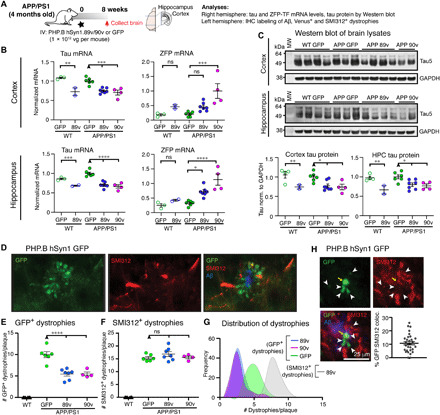
Fig. 7. Global tau reduction by AAV-PHP.B ZFP-TFs protects against Aβ plaque–induced dystrophies.

(A) Experimental overview: 4-month-old male APP/PS1 mice and age-matched control WT mice were retro-orbitally injected with a low dose (1 × 10¹² vg per mouse) of PHP.B hSyn1.89v, PHP.B hSyn1.90v, or PHP.B hSyn1.GFP, and the brain tissue was analyzed for RNA and protein content by immunohistochemistry (IHC) after 8 weeks. (B) Tau and ZFP-TF transcripts in the anterior cortex and hippocampus (HPC) of injected WT and APP/PS1 mice. Means ± SEM, one-way ANOVA, n = 2 to 7 mice per group. (C) Tau protein in cortical and hippocampal lysates of injected WT and APP/PS1 mice analyzed by Western blot. Tau protein is reduced by 20 to 30% in all mice injected with PHP.B hSyn1.89v or 90v. Means ± SEM, two-way ANOVA with Sidak’s test, n = 2 to 7 mice per group. (D) GFP (green) and axonal phospho-neurofilament (SMI312, red)–containing dystrophies around cortical Aβ plaques (blue) in a PHP.B hSyn1.GFP–injected animal. (E) The number of GFP+ dystrophies (immunolabeled with anti-GFP antibodies) per cortical plaque is reduced by ~50% in 89v- and 90v- compared to GFP-expressing APP/PS1 mice. Means ± SEM, two-way ANOVA with Sidak’s test, n = 4 to 7 mice per group, four to five cortical sections per mouse. (F) The number of SMI312+ dystrophies per cortical plaque is similar in 89v- and 90v- compared to GFP-expressing APP/PS1 mice. Means ± SEM, two-way ANOVA with Sidak’s test, n = 4 to 7 mice per group and four to five cortical sections per mouse. (G) Distribution of dystrophy numbers per cortical plaques in all mice and sections [same data as in (E) and (F)]. (H) Colocalization analysis of GFP-filled (green) and SMI312-filled (red) dystrophies around Aβ plaques (blue) in PHP.B hSyn1.GFP–injected APP/PS1 mice reveals that only ~10% of SMI312+ dystrophies originated from neurons transduced with the AAV and expressing GFP. Means ± SEM, n = 6 mice. MW, molecular weight.
