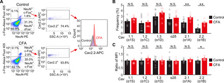
Fig. 8. Increase N-type Ca2+ channel expression in S1 with the pain induction.

(A) A representative example of FACS pseudo-color plot. Removal of doublets and debris was performed. Each experiment was obtained from 105 cells. Left: Neurons are gated from non-neurons on the basis of the expression of NeuN-PE, and active neurons are gated from inactive neurons on the basis of the expression of c-Fos–Alexa Fluor 405. Middle: Cav2.2 (α1B)–APC (voltage-dependent N-type Ca2+ channel)–positive neurons are gated/counted in active neurons in control and inflammatory pain model of CFA injection. Right: Histogram of expression of Cav2.2 (α1B) in NeuN, c-Fos, and Cav2.2 (α1B) triple-positive neurons in both models (black, control; red, CFA). (B) Frequency of NeuN, c-Fos, and each ion channel triple-positive neurons in various calcium ion channels in cells gated with (A) (black, control; red, CFA; n = 9 mice for each; **P < 0.01, paired t test. Error bars show means ± SEM.). (C) The ratio of the MFI of each Ca2+ channel in NeuN, c-Fos double-positive neurons in inflammatory pain model compared with control (black, control; red; CFA; n = 9 mice for each; *P < 0.05, paired t test. Error bars show means ± SEM.).
