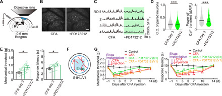
Fig. 9. Topical administration of N-type Ca2+ channel antagonist improved pain threshold.

(A) Schematic diagram showing method of the intraventricular PD173212 (N-type Ca2+ channel antagonist) administration and in vivo imaging. (B) Typical in vivo two-photon images of before and after a single dose of PD173212 in inflammatory pain model mice (3 days after CFA injection). Same neurons traced. Scale bar, 100 μm. (C) Calcium traces of typical four neurons. Each neuronal activity was traced before and after administration of PD173212. (D) Change in paired neuronal C.C., pCa2+ of neurons (n = 4295pairs, 203 neurons per five mice) in before and after a single dose of PD173212 in inflammatory pain model. ***P < 0.001, paired t test. Violin plots show median (black lines) and distribution of the data. (E) Change in pain threshold before and after a single dose of PD173212 in inflammatory pain model (n = 7 mice). *P < 0.05, paired t test. Error bars show means ± SEM. (F) Schematic diagram showing method of administration of the drug-soaked Elvax (PD173212; 2.5 mM) to S1HL through an open-skull cranial window. (G) Change in pain threshold by chronic local administration of PD173212 to S1HL/V1 [black, control; red, CFA; green, CFA + PD173212 (S1HL); orange, CFA + PD173212 (V1); *P < 0.05, **P < 0.01, and ***P < 0.001; two-way ANOVA followed by Bonferroni test, versus control]. Error bars show means ± SEM.
