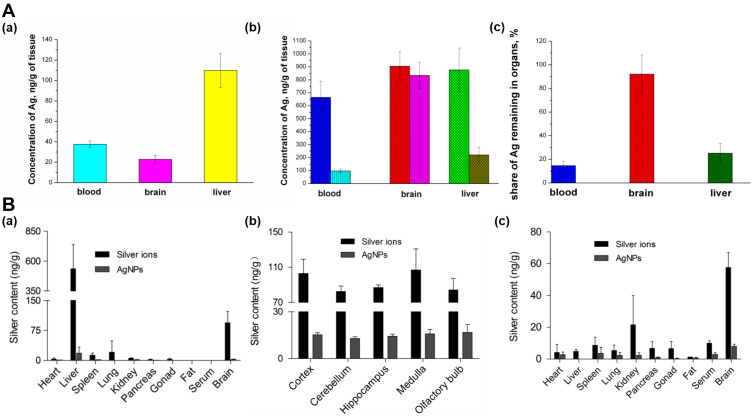
Figure 2.
Biodistribution and toxicity of SNPs. (A) Silver concentrations in different organs (a) after one-time exposure to SNPs of size 34 nm, stabilized with PVP (b) after long-time exposure, (c) Fractions of silver remaining in mouse organs after 1 month of washing up. Adapted with permission from. Antsiferova A, Buzulukov Y, Demin V, Kashkarov P,Kovalchuk M, Petritskaya E. Extremely low level of Ag nanoparticle excretion from mice brain in in vivo experiments. IOPConference Series. 2015;98:012003. Creative Commons Attribution 3.0 licence (https://creativecommons.org/licenses/by/3.0/).27 (B) Silver concentrations in main rat organs with daily intranasal administration of silver ions and SNPs of size 26 nm, at a dosage of 0.1mg kg−1 body weight day 1 (n = 4). (a) 4-week exposure (b) Silver distribution in different rat brain regions after 4-week exposure. (c) 12-week exposure. Adapted with permission from Wen R, Yang X, Hu L, Sun C, Zhou Q, Jiang G. Brain-targeteddistribution and high retention of silver by chronic intranasalinstillation of silver nanoparticles and ions in Sprague-Dawley rats. J Appl Toxicol. 2016;36(3):445–453. Copyright 2016 John Wiley and Sons.51

