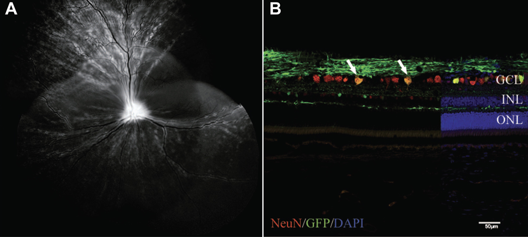
Figure 4.
Transduction of canine RGC following intravitreal injection of capsid mutated AAV2 (triple Y-F + T-V) with GFP transgene. (A) Representative confocal scanning laser ophthalmoscopy image obtained at 5 weeks post injection demonstrated widespread GFP fluorescence. (B) Immunohistochemical labeling of retinal cryosection with neuronal nuclei (NeuN) antibody (red) to label RGCs demonstrates a high number of cells colabeling with GFP (green). Cell nuclei are shown in blue with DAPI.
DAPI, 4’,6-diamidino-2-phenylindole; GCL, ganglion cell layer; GFP, green fluorescent protein; INL, inner nuclear layer; ONL, outer nuclear layer; triple Y-F+T-V, AAV2 based capsid was mutated by substitution of three surface-exposed capsid tyrosine (Y) residues with phenylalanine (F) and one threonine (T) residue with valine (V). Scale bar, 50 μm. Boyd et al. 2016, with permission.94

