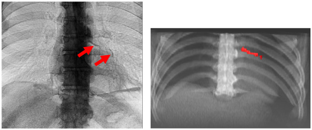
Fig 2.

Dual energy x-ray showing coronary calcification with corresponding corroborative CT (left and right, respectively). The calcification in the LVR has an Agatston score of 632. It is clearly present in dual energy chest x-ray image after processing with CorCalDx-viz. The CT image volume was registered with DE data to help determine correspondence. This is a high dose image acquisition.
