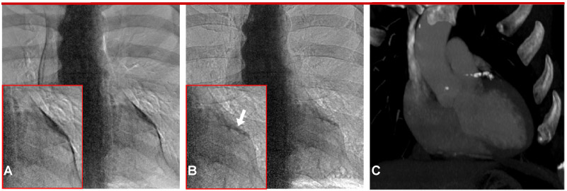
Fig 3.

CorCalDx-viz (B) compared to commercial “bone image” processing (A). The motion artifact present in A obscures the calcification and leads to confounding structures due to mismatched pulmonary arteries. Artifacts are greatly reduced in B. The corresponding CT calcium score image is shown in C with an Agatston score of 341 in the LAD. In this case, the CT image was not registered to the dual energy images in order to better show the extent of the calcification.
