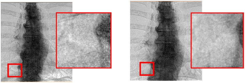
Fig 4.

Effect of conventional and high exposure acquisition (left and right, respectively). Images are acquired with conventional “right lung” and “spine + right lung” dose sensing chamber acquisitions, respectively. Clearly, inset images show that the higher dose processed images have much lower noise in the spine and lower portions of the image. The acquisition (kVp, mAs, ms) values for low and high voltage acquisitions are [(60 kVp, 250 mA, 14ms), (120 kVp, 200mA, 5ms)] and [(60 kVp, 630 mA, 9ms), (120kVp, 400mA, 3ms)], respectively. Averaging across patients, we determined that the conventional and high exposure acquisitions gave dose areas products of 2 dGy·cm2 and 3.5 dGy·cm2, respectively, as determined from the DICOM header. Nominally, we increases the dose area product by a factor of 1.75, from an average of 2 dGy·cm2 to 3.5 dGy·cm2. It was necessary to increase mA to maintain short exposures needed to minimize blurring.
