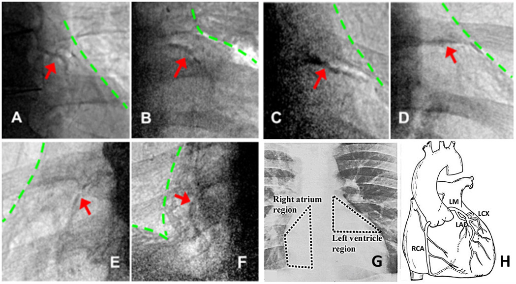
Fig 5.

Examples of coronary calcium found with CorCalDx-Viz. The red arrows indicate locations of coronary calcium plaques and the green lines indicate heart and diaphragm boundaries. (A)-(D) LM/LAD calcifications in left ventricle, the coronary artery calcium triangle. (E)-(F) RCA calcifications on the right atrium. (G)-(H) Anatomical illustration of regions important for coronary calcium, provided courtesy of reference [26].
